
Last Updated on June 15, 2026
Melanoma often looks like an unusual mole or dark spot that changes in size, shape, color, or texture over time. Common warning signs include asymmetry, irregular borders, multiple colors, a diameter larger than 6 millimeters, and evolution or noticeable changes. Melanoma may appear black, brown, tan, red, pink, blue, or even skin-colored. Early detection significantly improves treatment outcomes, making regular skin checks essential.
Melanoma is one of the most serious forms of skin cancer. Yet many people miss its earliest warning signs because it can resemble an ordinary mole, freckle, or age spot.
That similarity creates a dangerous problem.
A harmless-looking mark may actually be a growing cancer. By the time obvious symptoms appear, melanoma may have already spread beyond the skin.
The good news is that melanoma often provides visual clues long before it becomes advanced. Learning how to recognize those clues can make a life-changing difference.
In this guide, you’ll learn exactly what melanoma looks like, how it differs from normal moles, the warning signs dermatologists watch for, and the appearance of different melanoma types.
What Does Melanoma Look Like?
Melanoma usually appears as a new spot on the skin or as a change in an existing mole.
Unlike common moles, melanoma often has an unusual appearance. It may show several colors, irregular borders, or a shape that doesn’t match surrounding moles.
Some melanomas appear dark brown or black. Others may look red, pink, purple, blue, gray, or even flesh-colored.
Common visual characteristics include:
- Uneven shape
- Ragged or blurred edges
- Multiple colors within one lesion
- Enlargement over time
- Raised or thickened areas
- New skin spots appearing in adulthood
- Surface changes such as crusting or bleeding
A melanoma lesion may look dramatically different from the rest of your moles.
Dermatologists often call this the “ugly duckling sign.”
If one mole stands out from all the others, it deserves attention.
Understanding Melanoma Skin Cancer
Melanoma develops in cells called melanocytes.
These cells produce melanin, the pigment responsible for skin, hair, and eye color.
When melanocytes undergo genetic damage, they can begin multiplying uncontrollably. This growth creates melanoma.
Unlike many other skin cancers, melanoma has a stronger tendency to spread into nearby tissue, lymph nodes, and distant organs.
That aggressive behavior makes early detection extremely important.
Why Melanoma Looks Different From Other Skin Conditions
Healthy skin cells grow in an organized pattern.
Cancer cells do not.
As melanoma expands, it often grows unevenly. Different groups of cells produce varying amounts of pigment. This creates color variation and irregular shapes.
As a result, melanoma frequently develops:
- Uneven pigmentation
- Jagged borders
- Mixed colors
- Asymmetrical growth
- Unpredictable surface changes
These visual abnormalities help distinguish melanoma from benign moles.
How Melanoma Develops in Melanocytes
The process usually begins with DNA damage.
Common causes include:
| Risk Factor | Impact on Melanocytes |
|---|---|
| UV radiation | Damages cellular DNA |
| Severe sunburns | Increases mutation risk |
| Tanning beds | Delivers concentrated UV exposure |
| Family history | Raises inherited risk |
| Fair skin | Provides less natural UV protection |
Over time, accumulated mutations can transform healthy melanocytes into cancerous cells.
The resulting lesion may remain confined to the skin initially. However, untreated melanoma can eventually invade deeper tissues.
What Does Early Melanoma Look Like?
Many people assume melanoma always appears as a large black growth.
That assumption is incorrect.
Early melanoma often begins subtly.
It may start as a small change in a mole you’ve had for years. In other cases, it appears as an entirely new spot.
Recognizing these early changes offers the best opportunity for successful treatment.
Early Visual Signs of Melanoma
Early melanoma commonly presents as:
- A flat or slightly raised spot
- An enlarging mole
- Uneven pigmentation
- Irregular edges
- A lesion that differs from nearby moles
Sometimes the change occurs gradually over months.
In other cases, progression happens surprisingly quickly.
Because growth patterns vary, dermatologists emphasize monitoring any evolving skin lesion.
Common Color Changes
Color variation is one of the strongest warning signs.
A normal mole usually displays one consistent color.
Melanoma often contains multiple shades.
Possible colors include:
- Black
- Dark brown
- Light brown
- Tan
- Red
- Pink
- White
- Blue
- Gray
Some lesions contain several colors simultaneously.
This patchwork appearance should never be ignored.
Texture and Surface Changes
Texture often changes as melanoma develops.
You may notice:
- Roughness
- Scaliness
- Thickening
- Crusting
- Elevation
- Ulceration
A previously smooth mole that suddenly becomes rough deserves medical evaluation.
Why Early Detection Matters
Melanoma diagnosed early remains highly treatable.
When cancer remains confined to the skin’s outer layers, surgical removal often achieves excellent outcomes.
Once melanoma spreads deeper into the skin or other parts of the body, treatment becomes more complex.
For that reason, recognizing early appearance changes can have a profound impact on long-term health.
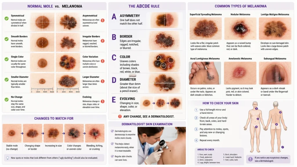
The ABCDE Rule for Identifying Melanoma
The ABCDE rule serves as one of the most valuable tools for spotting suspicious moles.
Dermatologists use this simple framework during skin examinations.
Each letter highlights a characteristic frequently seen in melanoma.
Asymmetry
A healthy mole often appears symmetrical.
If you draw an imaginary line through its center, both halves look similar.
Melanoma frequently breaks that pattern.
One side may appear larger, thicker, or shaped differently than the other.
Examples include:
- Uneven curves
- Distorted outlines
- Lopsided growth
Asymmetry remains one of the earliest warning signs.
Border Irregularity
Normal moles usually have smooth, clearly defined edges.
Melanoma often develops:
- Ragged borders
- Notched edges
- Blurred margins
- Uneven outlines
The border may appear as though it is spreading into surrounding skin.
Color Variation
One color is reassuring.
Several colors are concerning.
Melanoma commonly combines multiple shades within a single lesion.
Typical combinations include:
- Brown and black
- Black and blue
- Brown and red
- Pink and gray
The greater the color variation, the more suspicious the lesion becomes.
Diameter
Large size alone doesn’t confirm melanoma.
However, lesions larger than 6 millimeters deserve closer examination.
That’s roughly the size of a pencil eraser.
Some melanomas remain smaller than this threshold. Others grow significantly larger.
Size should always be evaluated alongside other warning signs.
Evolving Mole
Evolution may be the most important warning sign of all.
Any mole that changes over time requires attention.
Watch for:
- Growth
- Color shifts
- New symptoms
- Shape changes
- Texture changes
A changing mole tells an important story.
Your skin is signaling that something is happening beneath the surface.
Melanoma vs Normal Mole
At first glance, melanoma and a normal mole can look surprisingly similar.
That’s why many cases go unnoticed in the early stages.
The key difference is that healthy moles usually remain stable for years. Melanoma tends to change. It grows, evolves, and develops unusual characteristics that stand out from surrounding skin.
Key Visual Differences Between Melanoma and a Normal Mole
The table below highlights the most important distinctions.
| Feature | Normal Mole | Melanoma |
|---|---|---|
| Shape | Symmetrical | Often asymmetrical |
| Border | Smooth and even | Ragged, blurred, or irregular |
| Color | Usually one color | Multiple colors or shades |
| Size | Typically stable | Often enlarges over time |
| Evolution | Little to no change | Continues changing |
| Surface | Smooth | May become rough, crusty, or ulcerated |
| Symptoms | Usually symptom-free | May itch, bleed, or become tender |
A harmless mole tends to look predictable.
Melanoma rarely does.
The Ugly Duckling Sign
Dermatologists frequently use the “ugly duckling” concept.
Most people have moles that share a similar appearance. One mole may be darker, larger, or shaped differently than all the others.
That unusual mole is the ugly duckling.
For example:
- Most of your moles are small and round.
- One mole appears dark black and irregular.
- That mole deserves closer attention.
The ugly duckling sign often identifies melanoma before obvious ABCDE features appear.
When a Mole Becomes Suspicious
A mole becomes concerning when it starts behaving differently.
Pay attention if you notice:
- Sudden enlargement
- New color development
- Darkening
- Raised areas
- Bleeding
- Crusting
- Persistent itching
- Shape distortion
Many melanoma diagnoses begin with a patient noticing one small change.
Never assume changes are harmless simply because the lesion has existed for years.
Common Types of Melanoma and Their Appearance
Melanoma does not always look the same.
Several subtypes exist. Each has unique visual characteristics and growth patterns.
Understanding these differences can improve early recognition.
Superficial Spreading Melanoma
Superficial spreading melanoma is the most common form of melanoma.
It accounts for the majority of diagnosed cases.
What Does Superficial Spreading Melanoma Look Like?
It often appears as:
- A flat or slightly raised patch
- Irregular borders
- Multiple shades of brown and black
- Slow outward growth
Common colors include:
- Tan
- Brown
- Black
- Red
- Blue
- White
The lesion frequently expands across the skin surface before growing deeper.
Common Locations
Men often develop it on:
- Back
- Chest
- Shoulders
Women commonly develop it on:
- Legs
- Lower extremities
Nodular Melanoma
Nodular melanoma is one of the most aggressive melanoma types.
Unlike superficial spreading melanoma, it grows vertically into deeper tissues much earlier.
What Does Nodular Melanoma Look Like?
It often appears as:
- A dome-shaped bump
- A rapidly enlarging nodule
- A firm raised lesion
Colors may include:
- Black
- Dark brown
- Blue-black
- Red
- Pink
Because it grows quickly, nodular melanoma can become dangerous within months.
Warning Signs
Watch for:
- Rapid growth
- Bleeding
- Ulceration
- Elevated appearance
- Firm texture
Many patients describe it as a new lump that seems to enlarge week by week.
Lentigo Maligna Melanoma
This subtype usually develops after years of sun exposure.
It commonly affects older adults.
What Does Lentigo Maligna Melanoma Look Like?
Typical features include:
- Large flat patch
- Uneven pigmentation
- Irregular edges
- Gradual enlargement
Colors often include:
- Tan
- Brown
- Dark brown
- Black
The lesion may resemble an age spot initially.
Over time, darker areas emerge and the borders become increasingly irregular.
Common Locations
Frequently found on:
- Face
- Nose
- Ears
- Neck
- Forearms
Areas with decades of UV exposure face the highest risk.
Acral Lentiginous Melanoma
Acral lentiginous melanoma receives attention because it develops in areas that many people never inspect.
It can occur regardless of skin color.
What Does Acral Lentiginous Melanoma Look Like?
It may appear as:
- A dark patch
- A brown or black streak
- An enlarging pigmented area
The borders often become uneven over time.
Common Locations
This melanoma develops on:
- Palms
- Soles of the feet
- Fingernails
- Toenails
Many people discover it late because these locations are overlooked during skin examinations.
Amelanotic Melanoma
Amelanotic melanoma can be particularly difficult to identify.
Unlike most melanomas, it contains little or no pigment.
What Does Amelanotic Melanoma Look Like?
Instead of appearing dark, it may look:
- Pink
- Red
- Flesh-colored
- Slightly translucent
Many cases resemble:
- Pimples
- Rashes
- Insect bites
- Minor skin irritations
Because it lacks the classic dark appearance, diagnosis is sometimes delayed.
Why It Is Dangerous
People often expect melanoma to be black.
Amelanotic melanoma breaks that expectation.
Any persistent skin lesion that continues growing should be evaluated regardless of color.
What Does Melanoma Look Like on Different Body Parts?
Melanoma appearance varies depending on location.
Certain body areas also carry unique warning signs.
What Does Melanoma Look Like on the Face?
Facial melanoma often begins as:
- A slowly enlarging brown patch
- An irregular pigmented area
- A dark spot with uneven borders
It may resemble:
- Freckles
- Age spots
- Sun spots
Changes over time help distinguish melanoma from benign pigmentation.
What Does Melanoma Look Like on the Legs?
The legs represent one of the most common melanoma sites, particularly in women.
Warning signs include:
- Enlarging moles
- Multicolored lesions
- New dark spots
- Irregular skin patches
Many lesions develop on the lower legs.
Regular self-examinations help identify changes early.
What Does Melanoma Look Like on the Arms?
Arm melanomas frequently appear on sun-exposed skin.
Look for:
- Dark patches
- Asymmetrical moles
- Uneven pigmentation
- New growths
Long-term UV exposure contributes significantly to risk.
What Does Melanoma Look Like on the Back and Chest?
The back remains one of the most common melanoma locations in men.
Challenges include:
- Difficult visibility
- Delayed recognition
- Missed growth changes
Ask a partner or family member to examine hard-to-see areas regularly.
What Does Melanoma Look Like on the Scalp?
Scalp melanoma often remains hidden by hair.
Symptoms may include:
- Dark patches
- Raised bumps
- Bleeding areas
- Persistent sores
Because diagnosis often occurs later, scalp melanoma deserves special attention during skin checks.
What Does Melanoma Look Like Under a Fingernail?
Subungual melanoma develops beneath nails.
Common warning signs include:
- Brown streaks
- Black streaks
- Widening pigment bands
- Nail splitting
- Nail distortion
Nail Melanoma Warning Checklist
| Warning Sign | Why It Matters |
|---|---|
| Dark vertical streak | Common early feature |
| Widening pigment band | Suggests progression |
| Nail cracking | May indicate invasion |
| Pigment spreading to skin | Important melanoma clue |
| Persistent discoloration | Requires evaluation |
Many people mistake nail melanoma for bruising.
Unlike a bruise, melanoma does not gradually grow out with the nail.
Melanoma Symptoms Beyond Appearance
Visual changes often appear first.
However, melanoma can produce additional symptoms as it progresses.
These signs should never be ignored.
Itching Mole
Persistent itching may indicate activity within the lesion.
Cancer cells can trigger inflammation and irritation.
A mole that suddenly becomes itchy deserves attention, especially when combined with other ABCDE features.
Bleeding Mole
Bleeding without obvious injury is concerning.
Melanoma may:
- Crack
- Ulcerate
- Break down
These changes can cause spontaneous bleeding.
A mole should not repeatedly bleed on its own.
Crusting Lesion
Crusting often develops when surface tissue becomes damaged.
You may notice:
- Dry scales
- Repeated scabbing
- Flaking skin
If crusting continues for weeks, seek medical evaluation.
Painful Mole or Tender Skin Lesion
Pain is not always present.
When it occurs, it may indicate:
- Tissue invasion
- Inflammation
- Nerve involvement
Tender lesions warrant examination, especially if appearance changes accompany discomfort.
Non-Healing Sore
One of the most overlooked warning signs is a sore that refuses to heal.
Normal skin repairs itself.
Cancerous tissue often does not.
A persistent lesion that remains open for several weeks requires assessment.
What Increases the Risk of Melanoma?
Not everyone faces the same melanoma risk.
Certain factors significantly increase the likelihood of developing melanoma skin cancer.
UV Radiation Exposure
Ultraviolet radiation remains the leading environmental cause of melanoma.
Sources include:
- Sunlight
- Tanning beds
- UV lamps
Repeated exposure damages DNA within melanocytes.
Over time, mutations accumulate and cancer risk rises.
History of Severe Sunburns
Several blistering sunburns can substantially increase melanoma risk.
Childhood burns are especially important because skin cells remain highly vulnerable during early development.
Fair Skin
People with lighter skin often have:
- Less melanin
- Less natural UV protection
- Higher sensitivity to sun damage
Risk tends to be greater among individuals who:
- Burn easily
- Freckle frequently
- Have light-colored eyes
Large Numbers of Moles
The more moles present, the more opportunities exist for abnormal cellular changes.
Higher-risk groups include individuals with:
- More than 50 moles
- Dysplastic nevi
- Atypical moles
Monitoring becomes especially important for these individuals.
Family History
Genetics play an important role.
Risk increases when close relatives have been diagnosed with melanoma.
Inherited mutations may affect how the body repairs damaged DNA.
Weakened Immune System
A healthy immune system helps identify and destroy abnormal cells.
Reduced immune function may increase melanoma risk and affect disease progression.
Examples include:
- Organ transplant recipients
- Certain autoimmune conditions
- Long-term immunosuppressive therapy
Understanding risk factors helps explain why regular skin examinations are so important.
Early recognition remains one of the strongest defenses against melanoma.
How Dermatologists Diagnose Melanoma
Recognizing a suspicious lesion is only the first step.
A definitive melanoma diagnosis requires a professional evaluation. Dermatologists use specialized tools and techniques to determine whether a skin lesion is benign or cancerous.
The earlier melanoma is diagnosed, the better the outcome tends to be.
Skin Examination
A comprehensive skin examination often begins with a visual assessment.
The dermatologist evaluates:
- Size
- Shape
- Color
- Border characteristics
- Symmetry
- Evolution over time
They may also ask questions about:
- Recent changes
- Symptoms
- Family history
- Sun exposure
- Previous skin cancers
Many melanomas are discovered during routine skin checks before symptoms develop.
Dermoscopy
Dermoscopy is one of the most valuable tools in melanoma detection.
A dermoscope is a handheld device that magnifies the skin and provides enhanced visualization of structures beneath the surface.
This examination allows dermatologists to identify:
- Pigment patterns
- Vascular structures
- Border abnormalities
- Hidden color variations
Many melanoma features become visible only through dermoscopy.
Skin Biopsy
A biopsy remains the gold standard for diagnosis.
If a lesion appears suspicious, the dermatologist removes part or all of it for laboratory analysis.
Pathologists examine the tissue under a microscope to determine:
- Whether melanoma is present
- Tumor thickness
- Growth characteristics
- Degree of invasion
Biopsy results guide treatment decisions and staging.
Sentinel Lymph Node Evaluation
For thicker melanomas, doctors may evaluate nearby lymph nodes.
The sentinel lymph node is the first node likely to receive cancer cells if melanoma spreads.
Examining this node helps determine whether metastasis has occurred.
Melanoma Staging
After diagnosis, melanoma is assigned a stage.
Staging describes how far the cancer has progressed.
| Stage | Description |
|---|---|
| Stage 0 | Cancer confined to the outer skin layer |
| Stage I | Thin melanoma with limited invasion |
| Stage II | Thicker melanoma without lymph node spread |
| Stage III | Spread to nearby lymph nodes |
| Stage IV | Spread to distant organs |
Stage plays a major role in determining prognosis and treatment.
Melanoma Stages and Appearance Progression
Melanoma appearance often changes as the disease advances.
While every case differs, certain patterns are common.
Stage 0 Melanoma
Stage 0 is also called melanoma in situ.
Characteristics include:
- Flat appearance
- Localized growth
- Early color changes
- Limited skin involvement
At this stage, melanoma has not penetrated deeper tissues.
Stage I Melanoma
Stage I lesions often show:
- Irregular pigmentation
- Small enlargement
- Border abnormalities
- Mild asymmetry
Many patients notice only subtle visual changes.
Stage II Melanoma
As melanoma grows deeper, lesions may become:
- Larger
- Thicker
- Raised
- More irregular
Additional symptoms can appear.
These include itching and occasional bleeding.
Stage III Melanoma
At this stage, cancer has spread to nearby lymph nodes.
The original lesion may show:
- Ulceration
- Significant thickening
- Surface breakdown
Some patients notice enlarged lymph nodes.
Stage IV Melanoma
Advanced melanoma can spread to:
- Lungs
- Liver
- Brain
- Bones
The skin lesion itself may become highly irregular, ulcerated, and symptomatic.
Early detection aims to prevent progression to this stage.
Melanoma Treatment Options
Treatment depends on:
- Tumor thickness
- Cancer stage
- Location
- Overall health
Modern medicine has dramatically improved melanoma outcomes over the last decade.
Surgical Removal
Surgery remains the primary treatment for early-stage melanoma.
The procedure removes:
- The melanoma
- A margin of surrounding healthy tissue
Removing surrounding tissue reduces recurrence risk.
Many early melanomas require no additional treatment after successful surgery.
Sentinel Lymph Node Surgery
When melanoma reaches a certain thickness, nearby lymph nodes may be evaluated or removed.
This helps determine whether cancer has spread.
Immunotherapy
Immunotherapy has transformed melanoma treatment.
Rather than attacking cancer directly, these medications help the immune system recognize and destroy cancer cells.
Benefits include:
- Improved survival
- Long-term disease control
- Reduced tumor growth
Immunotherapy is frequently used for advanced melanoma.
Targeted Therapy
Some melanomas contain specific genetic mutations.
Targeted therapies focus on these mutations.
Benefits include:
- Precision treatment
- Rapid tumor response
- Improved disease control
Genetic testing helps identify eligible patients.
Radiation Therapy
Radiation uses high-energy beams to destroy cancer cells.
It may be used:
- After surgery
- For metastatic disease
- To reduce symptoms
Although not the primary treatment for most melanomas, it remains valuable in selected cases.
Treatment Comparison Table
| Treatment | Main Purpose |
|---|---|
| Surgery | Remove localized melanoma |
| Immunotherapy | Activate immune response |
| Targeted therapy | Attack specific mutations |
| Radiation | Control local disease |
| Lymph node surgery | Evaluate spread |
Melanoma Survival Rates and Prognosis
Many people immediately fear the worst after hearing the word melanoma.
Fortunately, survival rates improve dramatically when melanoma is found early.
Early-Stage Melanoma Survival
When melanoma remains confined to the skin, outcomes are often excellent.
Key advantages of early diagnosis include:
- Simpler treatment
- Lower recurrence risk
- Reduced spread
- Better quality of life
This is why skin checks matter so much.
Factors Affecting Prognosis
Several factors influence outcomes.
These include:
- Tumor thickness
- Ulceration
- Lymph node involvement
- Metastasis
- Overall health
Thinner melanomas generally carry better prognoses.
Why Early Detection Changes Everything
A tiny lesion discovered today may prevent a life-threatening cancer tomorrow.
Many successful melanoma treatments begin with a patient noticing a seemingly minor change.
That awareness saves lives.
How to Check Your Skin for Melanoma
Monthly self-examinations are one of the simplest ways to identify suspicious lesions.
You don’t need special equipment.
A good mirror and adequate lighting are usually enough.
The Monthly Skin Self-Exam Process
Inspect your:
- Face
- Neck
- Ears
- Chest
- Abdomen
- Back
- Arms
- Hands
- Legs
- Feet
- Scalp
- Nails
Many melanomas appear in unexpected places.
A complete examination matters.
Areas People Frequently Miss
Certain regions often go unchecked.
These include:
- Scalp
- Behind the ears
- Soles of the feet
- Between toes
- Under nails
- Lower back
Use mirrors or ask a trusted partner for assistance.
Self-Check Melanoma Checklist
Review every mole for:
- Asymmetry
- Border irregularity
- Color variation
- Diameter growth
- Evolution
Also watch for:
- Bleeding
- Itching
- Crusting
- Tenderness
- Non-healing sores
The Ugly Duckling Checklist
Ask yourself:
- Does one mole look different from all others?
- Has a spot recently changed?
- Does a lesion continue growing?
- Does it have multiple colors?
- Does it look unusual compared with nearby moles?
If the answer is yes, schedule an evaluation.
Melanoma Prevention Strategies
Although not all melanomas can be prevented, risk can be reduced significantly.
Protect Yourself From UV Radiation
Effective sun protection includes:
- Broad-spectrum sunscreen
- Protective clothing
- Wide-brimmed hats
- UV-blocking sunglasses
Consistent protection reduces cumulative skin damage.
Avoid Tanning Beds
Artificial tanning devices expose the skin to concentrated ultraviolet radiation.
Regular use significantly increases melanoma risk.
Perform Regular Skin Checks
Routine self-examinations improve the likelihood of detecting melanoma early.
Consistency matters more than perfection.
Schedule Dermatologist Visits
Professional skin examinations provide an additional layer of protection.
Individuals at higher risk may benefit from annual screenings.
Key Facts About Melanoma
Melanoma is the most dangerous form of skin cancer because it can spread to other parts of the body if not detected early.
The ABCDE rule remains one of the most effective methods for identifying suspicious skin lesions.
Early-stage melanoma is often highly treatable when diagnosed before it spreads.
Melanoma does not always appear black. Some lesions are pink, red, or skin-colored.
Regular skin self-examinations can help identify warning signs before symptoms become severe.
Frequently Asked Questions
What Does Melanoma Look Like in the Early Stages?
Early melanoma often appears as an unusual mole or spot with irregular shape, uneven color, or gradual growth. It may be flat initially and show subtle changes over time.
What Color Is Melanoma?
Melanoma can be:
- Black
- Brown
- Tan
- Red
- Pink
- Gray
- Blue
- White
- Skin-colored
Many lesions contain several colors simultaneously.
Can Melanoma Be Skin Colored?
Yes.
Amelanotic melanoma may appear pink, red, or flesh-colored because it contains little pigment.
What Does Melanoma Look Like Compared to a Normal Mole?
A normal mole is usually symmetrical with a consistent color and smooth border.
Melanoma often displays asymmetry, multiple colors, irregular edges, and ongoing changes.
Can Melanoma Look Like a Bruise?
Yes.
Certain melanomas may resemble bruises, especially under nails or in acral lentiginous melanoma.
Persistent discoloration that does not resolve should be evaluated.
How Fast Does Melanoma Grow?
Growth rates vary.
Some melanomas develop slowly over years.
Others, particularly nodular melanoma, may grow rapidly within weeks or months.
Does Melanoma Always Hurt?
No.
Many melanomas remain painless during early stages.
Pain is not required for diagnosis.
Can Melanoma Itch?
Yes.
Itching is a recognized warning sign, especially when accompanied by visual changes.
Is Every Changing Mole Melanoma?
No.
Many mole changes are benign.
However, every evolving mole deserves professional assessment.
Final Thoughts
So, what does melanoma look like?
The answer isn’t always simple because melanoma can take many forms. It may appear as an irregular mole, a dark patch, a rapidly growing bump, a nail streak, or even a pink skin-colored lesion. What connects these appearances is change. Melanoma tends to evolve, grow, and develop features that set it apart from ordinary moles.
The most important warning signs include asymmetry, border irregularity, color variation, increasing diameter, and evolution over time. Additional symptoms such as itching, bleeding, crusting, tenderness, and non-healing sores can provide further clues.
Learning how to identify melanoma does not replace professional medical evaluation. However, understanding what melanoma looks like can help you recognize suspicious lesions sooner.
When it comes to melanoma skin cancer, early detection remains one of the most powerful tools for protecting long-term health. A few minutes spent checking your skin each month could make a life-changing difference.

Sophia Bennett is a professional writer focused on explaining word meanings, expressions and everyday language in a simple and engaging way.